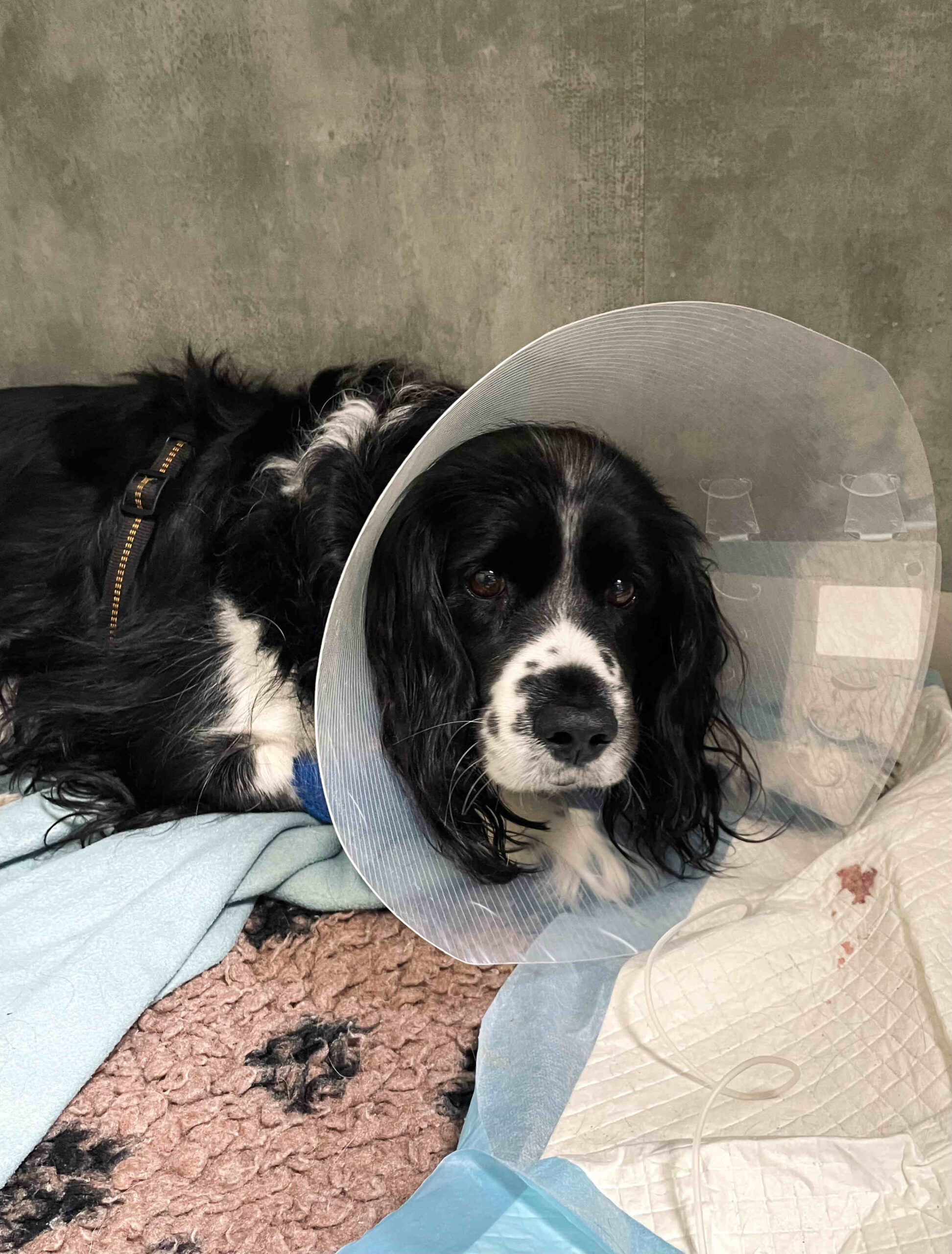
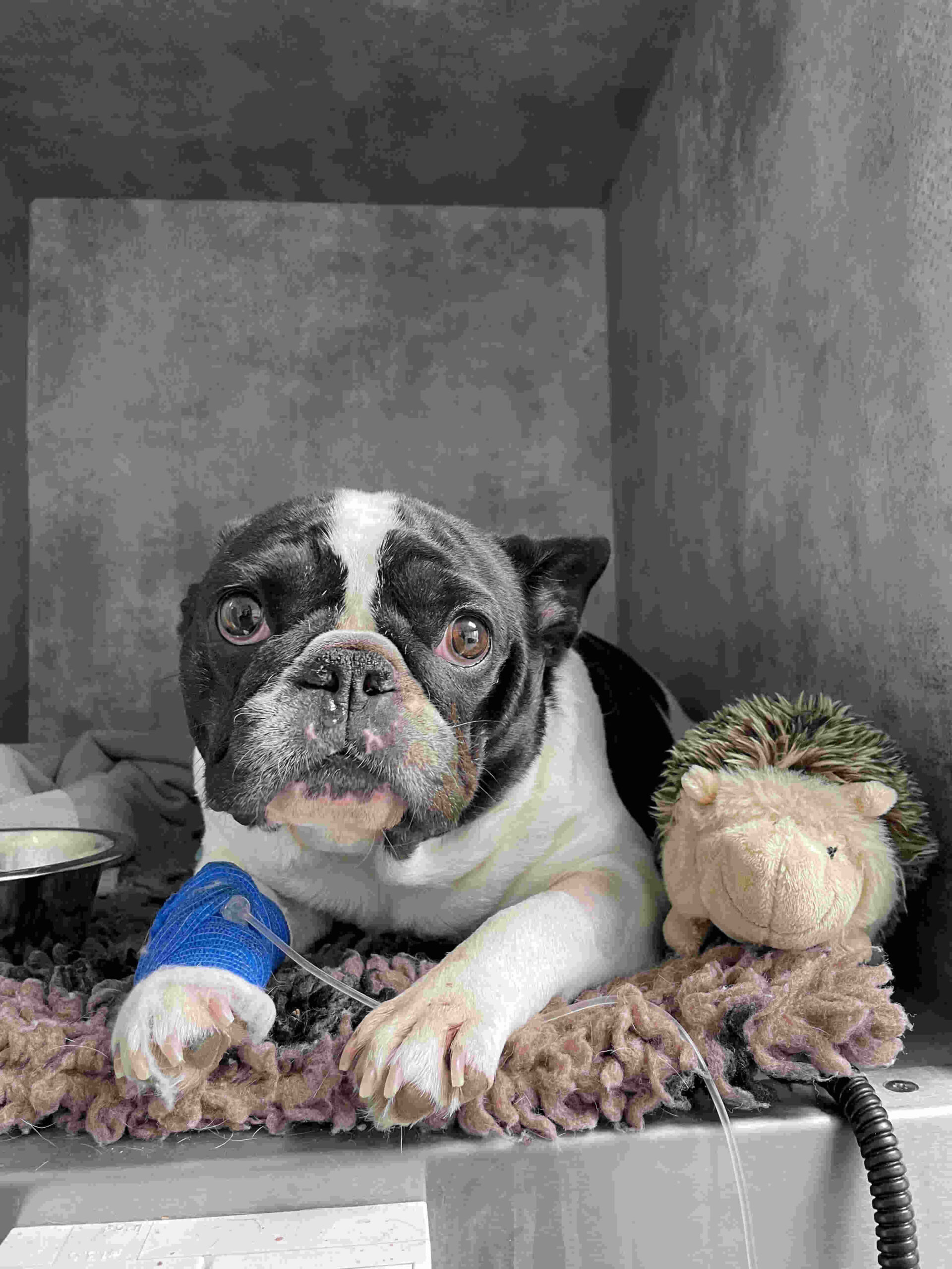
A diagnosis of an abdominal tumour always causes enormous anxiety for pet owners. Removal of the spleen in dogs, known medically as a splenectomy, is one of the most frequently performed surgical procedures in such cases. The operation involves the complete removal of the diseased organ, most often after the detection of suspicious tumours or extensive traumatic injury. It is worth stressing an extremely important and reassuring point right at the outset: an animal’s body has remarkable powers of adaptation, so a full, comfortable and long life for a dog without a spleen is entirely possible.
What does the spleen do in an animal’s body?
The spleen is a solid organ located in the front left part of the abdominal cavity. It performs several important functions in the animal’s body, mainly supporting the circulatory and immune systems. The organ acts as a kind of blood reservoir, releasing reserves of red blood cells at moments of sudden demand, for instance during intense exertion or severe stress. The spleen is also responsible for destroying old and damaged red blood cells, and it plays an active part in immune processes by producing specialised defence cells.
Important as these functions are, the spleen is not an organ that is absolutely essential for survival. After it is surgically removed, its tasks are largely taken over by other organs, primarily the liver and bone marrow. As a result, a patient who has come through surgery successfully and recovered properly returns to normal life.
Splenic tumours in dogs: the most common reason for surgery
The presence of abnormal structures on the surface of or inside the organ is the main reason a splenectomy is performed in dogs. These changes are usually detected during routine imaging or when the patient suddenly develops clinical signs.
Benign masses versus malignant tumours
Finding a mass on the spleen is not always a death sentence. Veterinary medicine distinguishes between benign growths and malignant tumours, and their appearance on ultrasound is often very similar.
A significant proportion of the masses diagnosed are benign. These include extensive haematomas resulting from injury, as well as benign nodular growths. In such cases, surgical removal of the organ is a definitive cure and completely eliminates the health problem.
On the other hand, extremely aggressive malignant tumours are also diagnosed. The most dangerous of these is haemangiosarcoma. It is a malignant tumour arising from the lining of blood vessels, characterised by very rapid growth and a strong tendency to spread, mainly to the liver and the right atrium of the heart.
It must be stressed that neither the ultrasound image nor the appearance of the tumour to the naked eye during surgery allows a benign mass to be definitively distinguished from a malignant one. The only reliable way to reach a final diagnosis is histopathological examination of a tissue sample taken immediately after the operation.
Breeds predisposed to splenic disease
Medical statistics clearly show that certain groups of dogs are far more likely to develop disease in this organ. The problem mostly affects older dogs, in whom the risk of cancer naturally rises with age. Large breeds are in the highest-risk group. Vets diagnose these conditions particularly often in:
- German Shepherds,
- Golden Retrievers,
- Labrador Retrievers.
Owners of these breeds should be especially vigilant and stick rigorously to a schedule of preventive imaging.
Rupture of a splenic tumour: an immediate threat to life
When a tumour or haematoma grows large, its walls become thin and extremely fragile. At that point, tissue damage is only a matter of time, and the tumour can rupture even during an ordinary walk or an exciting game. This is an absolutely critical moment. Blood pours from the damaged organ into the abdominal cavity at lightning speed, leading to profound haemorrhagic shock.
Owners need to know that the symptoms of a ruptured spleen in dogs are very sudden and dramatic. You may see:
- sudden and extreme weakness,
- porcelain-pale mucous membranes, visible on the gums,
- markedly rapid, shallow breathing,
- a rapidly swelling, tense and painful abdomen,
- complete loss of consciousness in extreme cases.
This condition requires immediate surgery at the nearest 24-hour veterinary clinic. In these situations, literally every minute counts.
Diagnosis: how are spleen problems detected?
Early detection is the key to successful treatment. The gold standard for diagnosing diseases of the internal organs is an abdominal ultrasound scan. Ultrasound allows the vet to assess the size and structure of the spleen accurately and to detect any abnormal masses within its tissue. This imaging is completely painless and non-invasive for the patient.
Detailed blood tests also play an extremely important role. An extended blood count makes it possible to assess any anaemia caused by blood loss, while a biochemistry profile gives a full picture of how the other organs are functioning before a planned anaesthetic. Having these tests done regularly as part of an annual check-up is the best investment in your dog’s health and safety.
What happens during a splenectomy at the veterinary clinic
From a medical standpoint, removing the organ is major abdominal surgery, requiring proper preparation and equipment. The procedure always begins with stabilising the patient. In emergencies involving massive internal bleeding, an urgent blood transfusion is often needed before the first incision or during the operation itself.
The operation is always performed under full general anaesthesia with intubation, with an anaesthetist continuously monitoring vital signs. The surgeon makes an incision along the midline of the abdomen, locates the diseased organ and then carefully ligates the numerous blood vessels in order to remove the entire spleen safely. It is worth emphasising that an operation performed on a planned basis, before the tumour ruptures, carries an incomparably lower risk of complications than life-saving surgery on a patient in profound shock.
Recovery: life for a dog after spleen removal
Post-operative care plays a key role in a safe recovery. For the first couple of weeks after discharge from the clinic, the most important thing is to strictly restrict the patient’s physical activity while the internal and external wounds heal. Walks should be very short and on the lead only, with no running or jumping.
The surgical wound must be physically protected from licking, which is usually achieved with a plastic protective collar or a special recovery suit. The attending vet will also prescribe appropriate medication for home, including strong painkillers and antibiotics to guard against infection. With proper aftercare, the patient soon regains strength, and a dog’s later life without a spleen is no different in quality from that of a fully healthy dog.
What is the prognosis after a splenectomy in dogs?
The patient’s future after successful surgery depends entirely on the results of the histopathological examination mentioned earlier. A frank assessment of the situation by the vet is crucial at this stage. If the laboratory confirms a benign mass, the prognosis is excellent: in this case, removing the diseased spleen means the patient is completely cured.
If, however, the microscopic examination confirms a malignant tumour, the prognosis for a splenic tumour in dogs becomes much more guarded and depends on how advanced the disease is. In the case of malignant haemangiosarcoma, surgery alone is very rarely enough. A veterinary oncologist will then discuss in detail with the owner the option of adjuvant chemotherapy. The aim of this treatment is to slow the progression of the disease, delay the onset of metastases and give the animal the longest possible life in good comfort.