A sudden change in the appearance of an animal’s eye is a situation that demands an immediate response from the owner. Glaucoma in dogs is an extremely dangerous, rapidly progressing neuropathy that leads to irreversible damage to the optic nerve and retina within a very short time. In veterinary medicine it is widely regarded as one of the most painful conditions a patient can suffer from. Understanding how it develops and recognising the early symptoms is the key to saving the animal’s sight and sparing it enormous suffering.
How glaucoma develops: what is aqueous humour?
To fully understand the nature of this condition, it helps to look at the basic anatomy and physiology of the eye. A healthy eye constantly produces a special fluid known in medical terminology as aqueous humour. This fluid nourishes the structures inside the eye, such as the cornea and the lens, and maintains the correct pressure within the eyeball.
The process relies on a delicate balance. Aqueous humour is produced by the ciliary body and must then drain freely out of the eye through a specialised anatomical structure called the drainage angle (iridocorneal angle). The trouble starts when, for whatever reason, this drainage mechanism becomes blocked. Excess fluid begins to build up inside the eyeball, resulting in dangerously high intraocular pressure. This mounting physical pressure destroys the delicate retinal ganglion cells and optic nerve fibres, both mechanically and by cutting off their blood supply, leading to permanent blindness.
Primary versus secondary glaucoma: what causes high pressure in the eye?
Ophthalmological diagnosis distinguishes two main types of the disease, which differ in their aetiology, that is, the underlying cause of the blockage in aqueous humour outflow.
Primary glaucoma is a genetically determined defect involving a congenital malformation of the drainage angle or dysplasia of the pectinate ligaments. The narrow drainage cleft closes completely over time, triggering a sudden, acute attack of the disease. This problem mostly affects particular groups of pedigree dogs. Highly predisposed breeds include the Basset Hound, Beagle, Cocker Spaniel, Siberian Husky and Chow Chow. Primary glaucoma always affects both eyes, although the attacks in each eye may be months or even years apart.
Secondary glaucoma, on the other hand, is a direct consequence of another disease already present in the eyeball that mechanically blocks the fluid’s drainage pathways. The most common causes include acute uveitis (inflammation of the uveal tract), lens luxation blocking the pupil, advanced cataracts, extensive physical trauma and tumours within the eye.
Tonometry: how is the pressure in a dog’s eye measured?
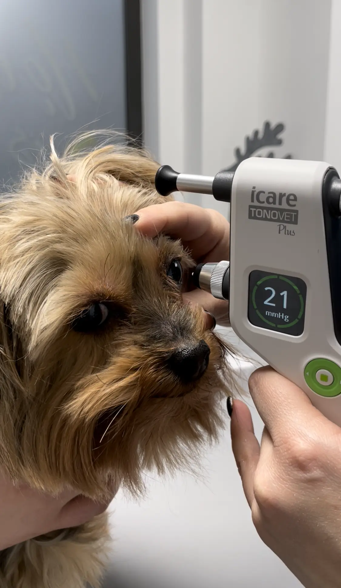
A key element of diagnosis and the absolute cornerstone of every ophthalmology visit is tonometry. This specialised test allows the pressure inside the eyeball to be measured accurately and objectively.
Modern veterinary clinics use electronic applanation or rebound tonometers for this purpose. The procedure is extremely quick, minimally invasive and, most importantly, completely painless for the patient. The device gently touches the surface of the cornea for a fraction of a second and reads the resistance offered by the eyeball. The test is so comfortable that in most cases it does not even require local anaesthetic drops.
An important rule of good veterinary practice is that the pressure in a dog’s eye is always measured in both eyes, even if clinically the problem appears to affect only one side. This makes it possible to catch early, symptom-free changes in the other eye and start preventive treatment.
Symptoms of glaucoma in dogs: signs that call for immediate action
With this disease, reaction time is absolutely decisive. Damage to the optic nerve can become irreversible within just a dozen or so hours of the first symptoms appearing. The clinical picture is divided into an acute and a chronic phase, each with slightly different warning signs.
Acute glaucoma attack: an emergency
An acute attack involves a sudden and extremely painful rise in pressure. In medicine this pain is compared to the worst migraine attacks in humans, combined with intense dull pressure inside the skull. The owner may notice the following symptoms of glaucoma in their dog:
- Severe eyelid spasm, known as blepharospasm: the animal keeps the eye squeezed shut and will not let you touch its face.
- Heavy, watery tearing.
- Marked congestion of the episcleral blood vessels: the white of the eye turns an intense, dark red.
- A fixed, widely dilated pupil that does not constrict at all, even in the bright light of a torch.
- A dramatic change in behaviour: sudden apathy, loss of appetite, hiding in dark places, whimpering and reluctance to do any physical activity.
Chronic glaucoma: the late stage
If the acute attack is not brought under control, the disease enters the chronic phase, in which the eyeball undergoes permanent structural changes. The dominant sign is a noticeable enlargement of the eyeball itself, known in medical terminology as buphthalmos. Corneal oedema also develops, which is why owners often report that their dog’s eye looks cloudy: its surface becomes hazy, bluish or completely milky. At this stage the animal is usually already permanently blind in the affected eye.
Treating glaucoma in dogs: medication and surgery
The choice of treatment depends on many factors, including the type of glaucoma, how advanced the changes are and whether the affected eye still has any visual reflexes. The overriding goal, however, is always to lower the pressure and eliminate the pain.
Medical treatment
The first step is emergency treatment based on aggressive drug therapy. This involves specialist drops to lower intraocular pressure, osmotic agents given intravenously and strong painkillers. Be aware, however, that medical treatment loses its effectiveness over time. The receptors in the eye become accustomed to the drugs, so the doses have to be increased and eventually the eye stops responding to medication altogether.
Surgical treatment of a sighted eye
If the patient reaches a specialist early enough and the eye can still see, microsurgical procedures are considered. One of the most advanced options is laser cyclophotocoagulation. This procedure uses a laser beam to precisely destroy part of the ciliary processes, point by point. Reducing the amount of tissue that produces aqueous humour directly translates into less fluid and a drop in intraocular pressure.
Treatment of a blind, painful eye
In patients where glaucoma has already caused complete and irreversible loss of sight, keeping the eyeball serves no therapeutic purpose and is merely a source of constant, crippling pain. In such cases the last resort, which from a medical point of view is the greatest relief for the animal, is radical surgery. This means either surgical enucleation, the complete removal of the eyeball with the eyelids sutured closed, or evisceration combined with the implantation of an intraocular silicone prosthesis, which preserves the natural appearance of the face.
A dog’s life without sight: is it the end of the world?
Understandably, the need to remove an eye or the irreversible loss of sight sounds like a death sentence to any owner and provokes strong emotional resistance. From the point of view of animal psychology and behaviour, however, the reality is quite different.
Unlike humans, animals rely far less on their sense of sight. Their perception of the world is based mainly on an excellent sense of smell and hearing. Once a painful, glaucomatous eye has been removed, the dog feels immediate relief. Most patients regain their old energy, appetite and playfulness within a few days of surgery. Animals are remarkably good at mapping their surroundings and can function perfectly normally, enjoying life free from chronic, exhausting head pain.