
-

Parasitic diseases in rabbits: the commonest threats and how to prevent them
Coccidiosis, encephalitozoonosis, ear mange and cheyletiellosis are the parasitic diseases pet rabbits meet most often, and they can go unnoticed for a long time. We explain how they are diagnosed in the laboratory, how they are treated, and which hygiene, quarantine and feeding rules keep infection at bay.
-

How to protect your dog’s paws in winter
Frost, ice and road salt leave a dog's pads irritated, cracked and sometimes frostbitten every winter. We explain how to pick a route and a protective wax before the walk, how to rinse and dry the paws properly once you are home, and which signs mean it is time to call the veterinary clinic.
-
Horner’s syndrome in dogs and cats: signs, causes, diagnosis and treatment
A drooping eyelid, a smaller pupil and a visible third eyelid on one side add up to Horner's syndrome. It is not painful in itself and does not harm sight, but it often signals another, more serious condition underneath. We explain the causes, the diagnostic steps, the pharmacological tests and the treatment options.
Foreign Bodies in Dogs and Cats: Symptoms, Types of Risk and the First Warning Signs
In veterinary medicine, a foreign body is any object or material that has found its way into an animal’s body when, physiologically, it should not be there. It may be something swallowed, something embedded in the soft tissues, or something migrating through the body. In clinical practice these cases are among the most common reasons for urgent consultations, because a foreign body in a dog or cat can lead to serious complications within a short time. This is especially true where it causes a gastrointestinal obstruction, compromises the blood supply to the bowel wall, perforates the bowel or leads to secondary infection of the tissues. The longer the object remains in the body, the greater the risk of necrosis, peritonitis, sepsis and a rapid deterioration in the patient’s condition.
A foreign body in the digestive tract: why does it need prompt assessment?
Whenever a dog or cat swallows a foreign body, prompt veterinary assessment is essential, even if the symptoms seem trivial at first. The patients most often seen in practice have eaten pieces of toys, socks, underwear, scraps of fabric, bones, bottle caps, string, ribbon or other everyday household items. Not every swallowed object causes a complete obstruction straight away, but the problem is that this cannot be safely judged from observation at home alone. The object may lodge in the oesophagus, stomach or intestine, and its clinical significance depends on its size, shape, elasticity, the sharpness of its edges and where exactly it has become wedged. Some patients initially show only mild signs resembling indigestion, but as the obstruction progresses the situation can quickly become an emergency.
It is also worth stressing that the danger does not come solely from the object being “stuck”. When a foreign body blocks the passage of food, it disrupts the workings of the digestive tract and can press on the wall of the organ until the blood supply is compromised. Under these conditions the tissues become starved of oxygen, weaker and more prone to damage. In advanced cases necrosis develops, followed by perforation, through which intestinal contents leak into the abdominal cavity. This is precisely why a foreign body in the stomach or intestines is not a minor incident but a problem that must always be assessed with great care.
Linear foreign bodies in cats and dogs: a particularly dangerous type of obstruction
A particularly dangerous variant is the linear foreign body in a cat or dog. This group includes all kinds of thread, string, tape, ribbon, tinsel, yarn and elastic bands. The problem is exceptionally common in cats, because their tongue is covered in backward-facing papillae that make it hard to spit out a thin material once it has caught. When a cat grabs a piece of thread or tinsel, swallowing is often involuntary and happens very quickly.
The danger lies in the fact that one end of the object can anchor itself, for instance at the base of the tongue, in the stomach or in the intestine, while the rest travels on along the digestive tract. The intestines try to move their contents onwards by peristalsis, but the anchored string does not move freely. As a result, the loops of bowel begin to bunch up and “thread” themselves onto the string. This is often compared to gathering fabric on a drawstring, and it is what produces the characteristic plication of the intestines. The taut material can then act like a fine blade, gradually cutting through the wall of the digestive tract. The consequences are perforation, leakage of intestinal contents into the abdomen and the development of peritonitis. A linear foreign body is therefore regarded as one of the more dangerous forms of gastrointestinal obstruction in small animals.
In practice, owners should remember one rule: if a thread, tape or ribbon is protruding from the animal’s mouth or anus, never pull it out yourself. Even a gentle tug can increase the tension on the anchored material and cause deep tissue damage. The only safe course of action is to get the animal to a veterinarian quickly, so that the situation can be assessed and the right approach chosen.
Migrating foreign bodies: grass seeds, splinters, thorns and needles
In veterinary medicine, a foreign body does not only mean something that has been swallowed. An equally important group is made up of migrating foreign bodies, that is, objects that pierce the skin or mucous membranes and then work their way deeper into the tissues. These include splinters, thorns, needles and grass seeds (grass awns). Grass seeds are especially treacherous, because their structure means they can effectively move in only one direction: ever deeper. They can enter the body through the nose, mouth, ear or eye, as well as through the skin, particularly around the paws. Over time they can migrate further, causing chronic inflammation and creating lesions that are slow to heal.
A grass seed in a dog’s ear is one of the classic seasonal problems seen after walks through meadows, roadside verges and tall, dry grass. Plant material of this kind can enter the ear canal and, with every movement of the head, travel deeper. Other common sites are the spaces between the toes, the conjunctival sac and the nasal cavity. If a grass seed lodges in the nose, violent bouts of sneezing, head shaking and a one-sided nasal discharge are typical. If it pierces a paw, the dog or cat may lick one spot intensively, limp or avoid putting weight on the limb. A foreign body in the eye, meanwhile, often causes marked squinting, watering and rapidly increasing swelling.
One point deserves clarification here: a migrating foreign body is not “contagious”, but it does carry a high risk of secondary infection. By carrying bacteria, fungi and environmental contaminants deep into the tissues, it can lead to abscesses, draining tracts and painful inflammation. The longer the material goes unnoticed, the harder it becomes to pinpoint its location. This is why, in cases of persistent discharge, swelling or recurring inflammation after a walk, a migrating foreign body should always be high on the list of diagnostic suspects.
What are the symptoms of a swallowed foreign body in dogs and cats?
The symptoms of a swallowed foreign body in dogs and cats depend above all on the type of object, where it has lodged and how much time has passed since the incident. In some animals the onset is sudden and very obvious; in others the signs develop gradually. The most common are vomiting, loss of appetite, lethargy, dehydration, abdominal pain and behavioural changes linked to discomfort. The animal may become apathetic, reluctant to move, hide away, react nervously when its belly is touched, or refuse food and water. With a partial obstruction the symptoms can come and go, which is misleading for owners and lulls them into a false sense of security.
Signs from the digestive tract are the most common. With a foreign body in the stomach or intestines, we see recurrent vomiting, regurgitation of food, excessive salivation, abdominal tenderness, bloating, lack of appetite, and difficulty passing stools or passing only small amounts. Some patients develop diarrhoea, sometimes with traces of blood. With a linear foreign body, an additional clue may be persistent pawing at the mouth or distress caused by a thread wrapped around the base of the tongue. Whatever the exact clinical picture, persistent vomiting, refusal to eat and obvious discomfort always call for an urgent consultation.
Signs of pain are also very important. An animal with an obstruction may adopt the so-called “prayer position”, stretching its front legs as far forward as possible with its hindquarters raised. This posture is not a diagnosis in itself, but it may point to severe abdominal pain and should catch the owner’s attention. Equally alarming are a tense abdomen, reluctance to lie down, whimpering when touched, and a marked change in behaviour when the animal is picked up or stroked around the belly.
Symptoms of migrating foreign bodies: what to look out for after a walk
The symptoms of migrating foreign bodies depend on the point of entry and the direction of migration. A grass seed in a dog’s or cat’s ear often presents suddenly: vigorous head shaking, tilting the head to one side, scratching at the ear and obvious distress. A foreign body in the nose usually causes fits of sneezing, rubbing the muzzle along the ground, snorting, and sometimes a one-sided discharge or bleeding. In the paw, the typical signs are lameness, persistent licking of one space between the toes, rapidly developing swelling and pain on touch. If the eye is affected, there may be marked squinting, watering and swelling. These signs are not exclusive to grass seeds, but after a walk through tall grass they should immediately raise that suspicion.
Diagnostic imaging: what does an X-ray not show?
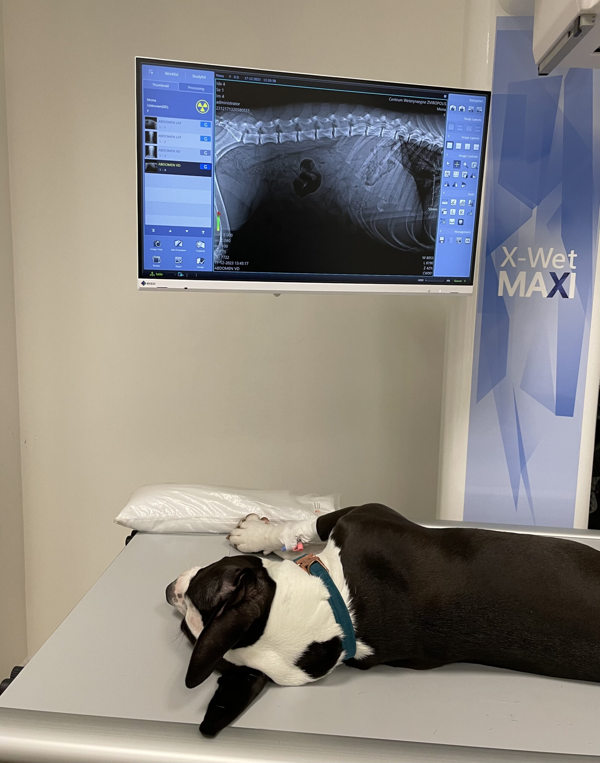
Confirming the presence of a foreign body in a dog or cat requires well-planned diagnostic imaging, because the history and clinical signs alone very often do not allow us to say for certain what we are dealing with. A conventional radiographic examination, or X-ray, is usually the first step in assessing a patient with a suspected gastrointestinal obstruction or a swallowed object. Radiography remains the basic screening test because it is widely available, quick to perform and helps determine whether the situation calls for continued conservative management or for urgent endoscopic or surgical intervention. In practice, well-taken radiographs help detect radiopaque foreign bodies such as metal items, some bones, stones and certain denser synthetic materials, but their value is not limited to simply “seeing” the object. Very often, assessing the indirect signs of obstruction is just as important: segmental dilation of bowel loops, an abnormal gas pattern, stacking of the intestines or abnormal contents in the lumen of the digestive tract.
It must be said clearly, however, that conventional X-rays have their limitations. Not all foreign bodies are radiopaque, so thin plastics, pieces of rubber, fabric, sponge, string and other materials with a density similar to soft tissue may not be directly visible on the image. In such cases the veterinarian looks primarily for indirect signs, meaning changes that point to a mechanical obstacle to the passage of food. This is why a normal or inconclusive X-ray does not yet rule out a foreign body. In clinical practice it means that if the symptoms persist, the patient often needs further investigation and sometimes repeat imaging. In selected situations a contrast study can help, as it outlines the lumen of the digestive tract more clearly and shows whether the contrast passes through normally or is held up or distorted by an obstruction.
An equally important, and in many cases decisive, examination is abdominal ultrasound. Ultrasound for animals is particularly useful when a foreign body that cannot be seen directly on an X-ray is suspected, or when the condition of the stomach and bowel wall needs to be assessed more closely. A major advantage of ultrasonography is that it allows soft-tissue structures to be assessed in real time, along with the motility of the digestive tract and intestinal peristalsis. In practice, ultrasound can help visualise both the obstruction itself and its consequences, such as thickening of the bowel wall, the presence of fluid, impaired motility or features suggesting a higher risk of tissue damage. Studies comparing radiography and ultrasonography indicate that in some cases it is ultrasound that confirms a gastrointestinal foreign body in small animals more effectively, especially when radiography gives no clear answer. For this reason the two methods usually do not compete but complement each other.
It is also worth adding that the choice of imaging depends on where the problem is suspected to be. If a foreign body in the oesophagus is suspected, it is not only abdominal images that matter but also appropriately chosen views of the chest. If, on the other hand, the veterinarian suspects a linear foreign body, its appearance may be subtler and require assessment of specific dilation patterns and an unusual distribution of gas within the bowel loops. This is another reason why the interpretation of imaging should always be weighed against the clinical signs, the findings on palpation and the patient’s overall condition. The technology does not replace clinical reasoning; it is an extension of it and a tool for making the right treatment decision.
Methods of removal: from inducing vomiting to soft-tissue surgery
The choice of method for removing a foreign body depends on several key factors: how much time has passed since the object was swallowed, what kind of object it is, where it is located, the extent of tissue damage and the animal’s overall clinical condition. There is no single universal procedure that suits every patient. That is why the approach has to be tailored to the individual, and the veterinarian’s aim is not only to remove the obstruction but also to limit the risk of perforation, aspiration, peritonitis and other complications. The sooner the patient reaches the clinic, the better the chances of using a less invasive method and getting the problem under control quickly.
One possible option in the early phase is inducing vomiting with medication, but it must be stressed that this is neither a routine procedure nor one that is entirely harmless to the patient. Inducing emesis can carry adverse effects such as prolonged vomiting, aspiration and, in some cases, even serious complications, so the risks and benefits must be weighed up every time. The exact time since ingestion, the nature of the object and the risk of damaging the oesophagus as the object comes back up all matter. In practice, vomiting is not induced after ingestion of sharp objects, corrosive substances, batteries, large objects that could lodge in the oesophagus, or linear foreign bodies. What is more, the way vomiting is induced differs between species: dogs are given different drugs from cats, so generic home “attempts to make the animal sick” are dangerous and should never take the place of a veterinarian’s decision.
If emptying the stomach with medication is not indicated, does not work, or the object is still in the oesophagus or stomach, endoscopy plays a very important role. Endoscopic removal of a foreign body involves passing a flexible endoscope, fitted with a camera and working instruments, through a natural body opening so that the object can be grasped and retrieved. This approach is enormously valuable because it allows the mucosa to be examined directly at the same time and does not require the abdomen to be opened surgically. Although the procedure is carried out under general anaesthesia, it is considered minimally invasive and often shortens the recovery period considerably. Endoscopy is used not only for foreign bodies in the oesophagus or stomach; depending on the equipment available and the location of the problem, it can also be used to remove material from the throat, nose or airways.
If, however, the foreign body has moved further into the intestines, caused a complete obstruction, become wedged in a way that rules out endoscopic retrieval, or caused perforation and tissue necrosis, the intervention of a veterinary surgeon becomes necessary. A laparotomy in a dog or cat involves surgically opening the abdominal cavity to locate the obstruction, assess the condition of the organs and remove the problem safely. Depending on the location, a gastrotomy is performed when the stomach needs to be opened, or an enterotomy when the obstruction is in the intestine. In the most severe cases it may be necessary to resect a section of bowel and join the healthy ends of the digestive tract back together. This is a more demanding procedure than endoscopy, but it can be the only chance of saving the patient where there is advanced necrosis, perforation or generalised inflammation within the abdomen. After such surgery, intensive postoperative care, fluid monitoring, pain management and appropriately chosen medication are essential.
It is worth remembering that treatment for a foreign body does not end the moment the object is removed. The prognosis also depends on how long the object remained in the body, whether the wall of the digestive tract was damaged, and whether electrolyte imbalances, dehydration, infection or sepsis developed. This is why early recognition and a well-chosen method of removal are so clinically important. In many cases the success of treatment is decided not by the hours spent in the operating theatre, but by how quickly the diagnostic and treatment decisions were made at the very start of the process.